
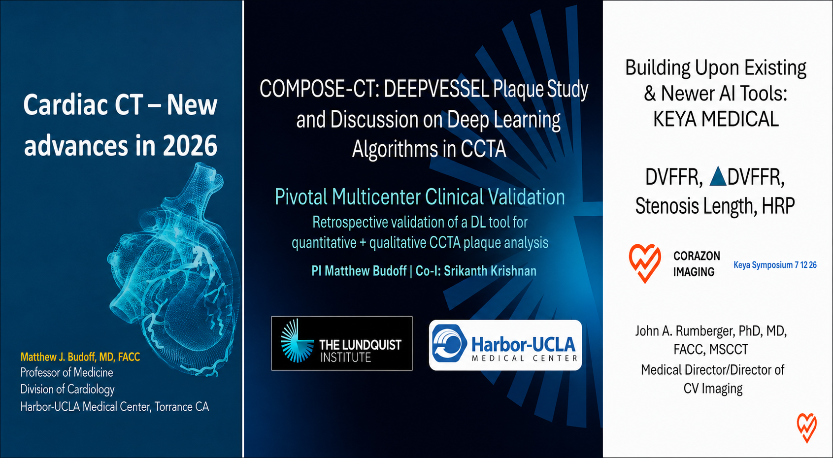
Keya Medical Sponsored Webinar at SCCT 2026
Coronary CT angiography (CCTA) continues to evolve from a test focused primarily on stenosis into a more comprehensive platform for evaluating coronary artery disease. When CCTA is combined with quantitative plaque analysis and CT-derived fractional flow reserve (CT-FFR), clinicians can assess coronary anatomy, atherosclerotic disease burden, and the probable physiological significance of individual lesions.
During Keya Medical’s sponsored webinar at SCCT 2026, experts in cardiac CT discussed advances in comprehensive CCTA assessment, presented results from the COMPOSE-CT clinical validation study, and examined how plaque and physiological findings can be interpreted together in clinical practice.
Expanding the clinical value of CCTA
Dr. Matthew Budoff moderated the program and reviewed how advances in cardiac CT have improved image quality, shortened breath-hold times, reduced contrast and radiation requirements, and increased clinical throughput.
He emphasized that contemporary CCTA can provide substantially more information than the degree of luminal narrowing alone. It can help clinicians:
- Detect obstructive and nonobstructive coronary disease
- Identify and characterize coronary plaque
- Quantify overall atherosclerotic disease burden
- Support more individualized risk assessment
- Inform preventive treatment decisions
- Evaluate the probable physiological significance of coronary lesions with CT-FFR
A central theme of the webinar was the importance of evaluating plaque burden and composition alongside stenosis severity. Quantitative plaque information may help clinicians identify patients who could benefit from more intensive preventive therapy—even when their disease is not producing severe anatomical obstruction.

Dr. Srikanth Krishnan presented findings from COMPOSE-CT, the multicenter clinical performance study supporting DEEPVESSEL Plaque.
The retrospective study evaluated 147 CCTA examinations and 441 coronary vessels. The study dataset represented a range of patient demographics, disease burden, stenosis severity, and CT scanner vendors. Importantly, the validation cases had not been used to train or develop the algorithm.
DEEPVESSEL Plaque results were compared with manual plaque annotations established through consensus among three SCCT Level III readers. The algorithm and expert-reader assessments were blinded, and the study’s performance criteria were established before the analysis.

The study also reported strong agreement for stenosis categorization and strong lumen and vessel-wall segmentation performance. According to the presentation, the algorithm exceeded all prespecified performance criteria.
The faculty also discussed important study limitations. COMPOSE-CT was retrospective, and its participating clinical sites were geographically concentrated in the Los Angeles area. Expert-reader consensus served as the reference standard, rather than an independent imaging or pathological modality such as intravascular ultrasound or histology.
Looking beyond a single CT-FFR value
Dr. John Rumberger discussed the interpretation of DEEPVESSEL FFR and explained why clinicians should consider the full pressure pattern along a coronary vessel rather than relying exclusively on a single distal CT-FFR value.
A distal value provides important information, but it does not always reveal whether a pressure decline is caused by a discrete focal lesion or by diffuse disease. This distinction can be especially important in borderline cases.
The interpretation described during the webinar considered several complementary factors:
- Distal CT-FFR value
- Magnitude of the pressure change across a lesion
- Location of the pressure drop
- Lesion length
- Focal versus diffuse disease
- Stenosis severity
- Plaque volume and composition
- Patient symptoms and overall clinical context
A relatively abrupt focal pressure drop may identify a lesion with greater physiological significance, while a gradual decline along the vessel may be more consistent with diffuse atherosclerotic disease. The panel emphasized that these patterns should inform clinical interpretation rather than be treated as automatic thresholds for intervention.
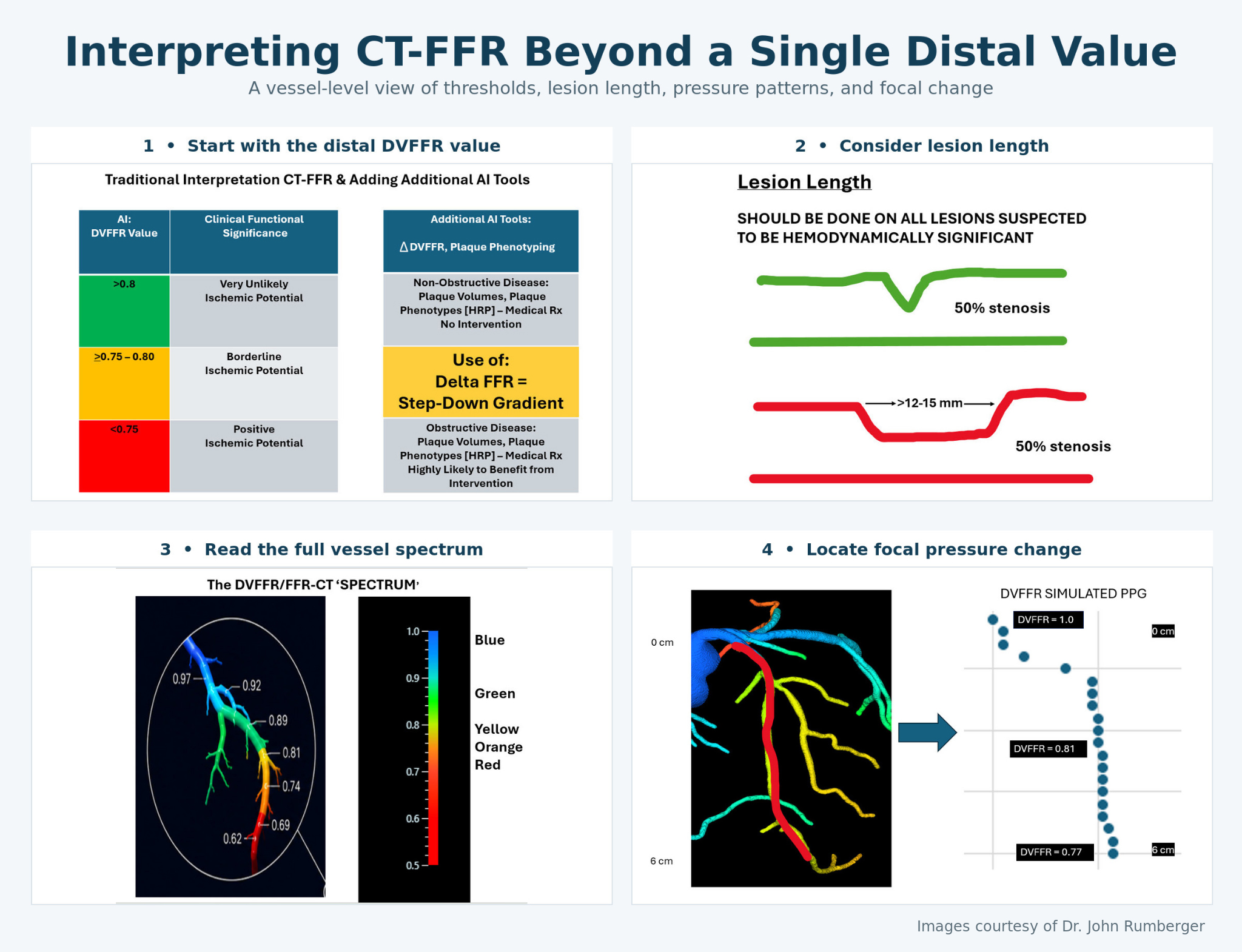
Beyond a single distal CT-FFR value: Interpreting the full pressure pattern—including focal pressure change, lesion length, and focal versus diffuse disease—can provide a more complete understanding when considered alongside stenosis severity, plaque characteristics, symptoms, and the broader clinical context.
Combining anatomy, plaque, and physiology
- Plaque analysis and CT-FFR provide different but complementary information:
- Plaque analysis describes the extent and composition of coronary atherosclerosis.
- Stenosis analysis describes the degree of anatomical narrowing.
- CT-FFR estimates the probable effect of coronary disease on blood flow.
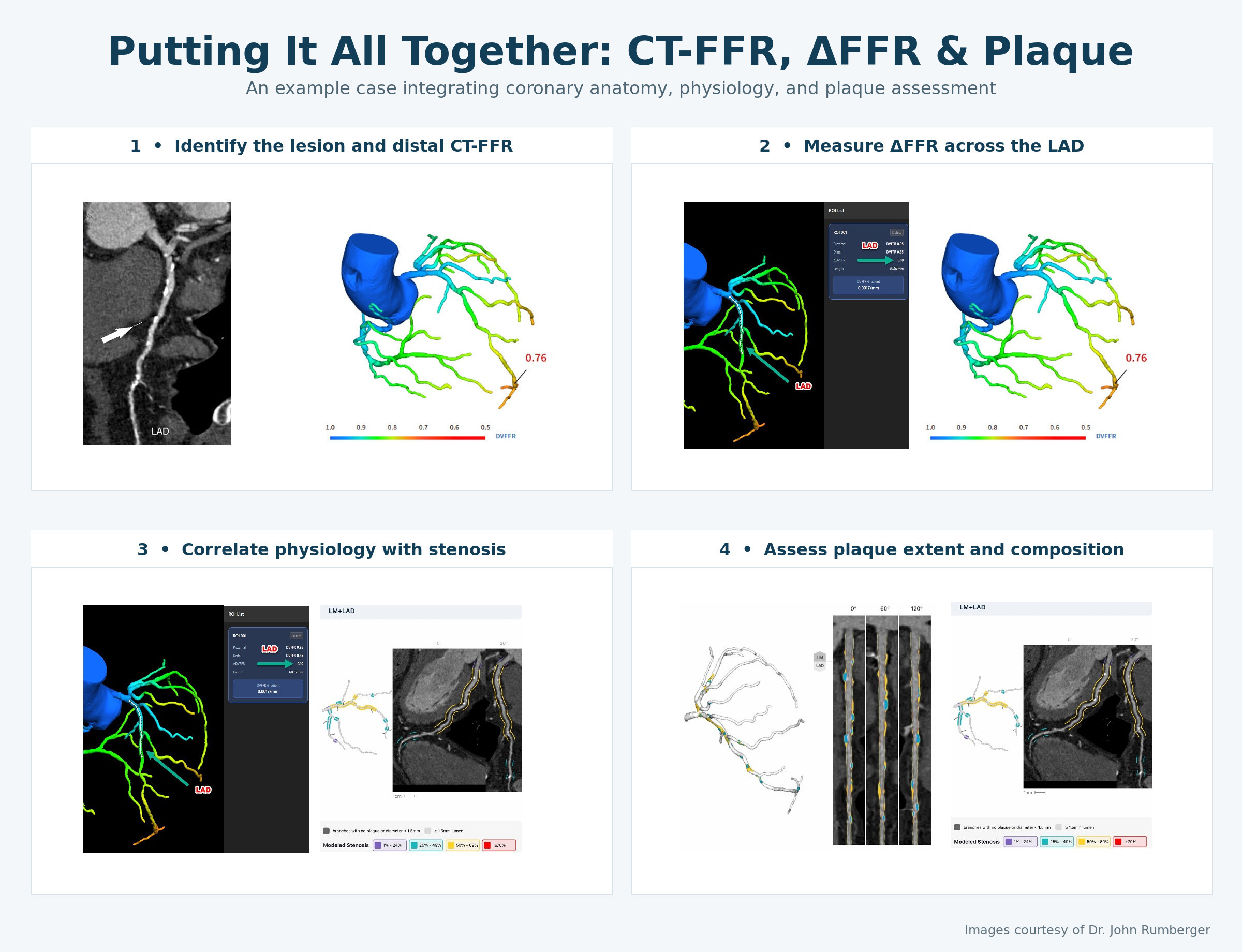
When these findings are concordant, the clinical interpretation may be relatively straightforward. When they differ—or when measurements fall within borderline ranges—physician judgment becomes even more important.
The panel described the clinician as the “interpreter of the interpretation.” AI-assisted measurements can provide detailed and reproducible quantitative information, but the physician must integrate those findings with symptoms, clinical history, risk factors, and the patient’s broader cardiovascular profile.
Practical considerations discussed by the faculty
The panel also addressed several common clinical questions.
Dense coronary calcification
The COMPOSE-CT study did not exclude patients according to a specific calcium-score threshold. Expert readers adjusted image windowing and evaluated pixel transitions to define lumen and vessel boundaries where possible. Nevertheless, extensive calcification remains challenging and may particularly affect specificity.
Patients with prior stents
CT-FFR is not currently applied through stented segments in the same manner as native coronary vessels. CCTA may still be used to assess the stent for restenosis, while eligible nonstented vessels may be evaluated with CT-FFR. Plaque analysis can also provide information about disease elsewhere in the coronary tree.
Longitudinal follow-up
The faculty discussed the potential role of serial imaging in monitoring disease progression or regression. Depending on the patient’s clinical risk and initial findings, repeat assessment over an interval of approximately two to five years may provide useful information.
Toward a more comprehensive CCTA assessment
The webinar highlighted the growing potential of CCTA to answer several clinically important questions within a single diagnostic pathway:
- Is coronary atherosclerosis present?
- How much plaque is present?
- What type of plaque is present?
- How severe is the anatomical narrowing?
- Is the disease likely to impair coronary blood flow?
COMPOSE-CT demonstrated strong agreement between DEEPVESSEL Plaque and the consensus of expert readers for plaque-volume and stenosis assessments. The broader clinical discussion showed how plaque quantification and CT-FFR can add complementary information to the original CCTA examination.
These technologies are intended to support—not replace—clinical judgment. Their greatest potential lies in helping clinicians integrate anatomy, plaque burden, plaque composition, physiology, symptoms, and patient risk into a more comprehensive assessment of coronary artery disease.