

Beyond Stenosis: How Quantitative CCTA Brings Plaque and Physiology Into Focus
Clinical Perspectives on Quantitative CCTA and a First Look at the DVCloud Interactive Workflow
Coronary CT angiography has moved well beyond a narrow assessment of luminal stenosis. Advances in scanner technology, image reconstruction, and artificial intelligence now allow clinicians to examine coronary anatomy, plaque burden and composition, and the probable physiological effect of coronary disease within a more comprehensive CCTA pathway.
During Keya Medical’s sponsored webinar at SCCT 2026, Dr. Irfan Shafique, Dr. Armin A. Zadeh, and Dr. Matthew J. Budoff reviewed the current evidence, practical applications, and remaining questions surrounding quantitative CCTA. Their presentations offered complementary perspectives: the rapid expansion of imaging biomarkers, the need for rigorous evidence and standardization, and the clinical value of identifying and treating coronary disease earlier.
The role of biomarkers and CT FFR in the assessment of cardiovascular disease
Dr. Shafique opened with an overview of the imaging biomarkers that can now be derived from cardiac CT. These include CT-derived fractional flow reserve, total plaque volume, percent atheroma volume, plaque composition, coronary calcium, and perivascular fat attenuation. Together, these measures can provide information about the presence, extent, and characteristics of coronary disease that may not be captured by stenosis severity alone.
He also discussed how AI is being applied throughout the CCTA workflow, from motion correction and image reconstruction to coronary plaque analysis and CT-FFR. These developments can improve reproducibility and help clinicians evaluate complex datasets more efficiently, but image quality and appropriate clinical interpretation remain essential.
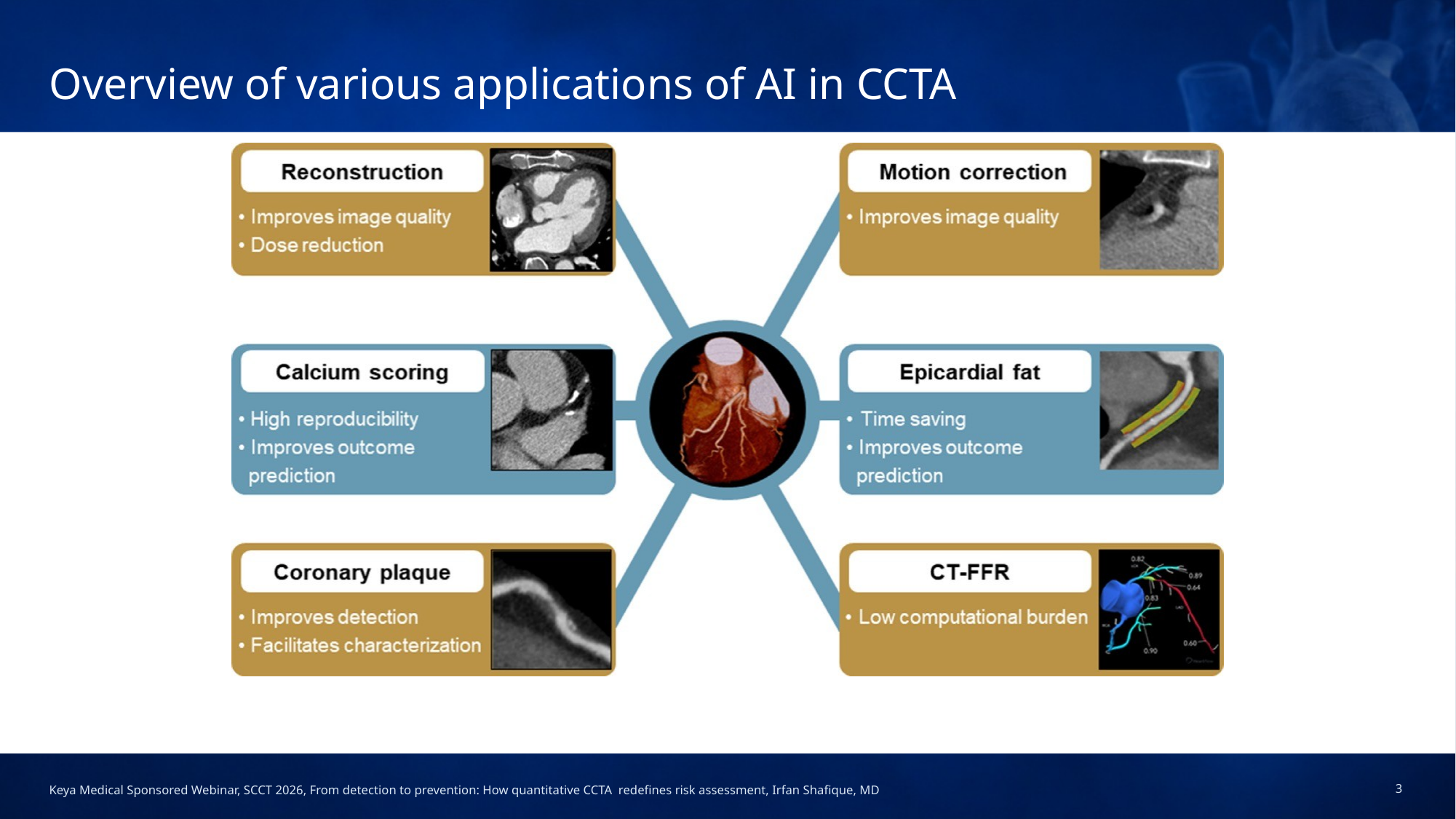
Applications of AI across the CCTA workflow. Source: presentation by Irfan Shafique, MD.
An anatomical narrowing does not always produce a corresponding reduction in blood flow. CT-FFR adds physiological context by estimating whether coronary disease is likely to be hemodynamically significant. Dr. Shafique reviewed evidence supporting CT-FFR as a noninvasive gatekeeper that can help reduce unnecessary invasive coronary angiography and improve the selection of patients who may benefit from further evaluation.
His presentation included validation data for DEEPVESSEL FFR. The ADAPT study formed the basis for the product’s 2022 FDA clearance, with reported vessel-level accuracy of 86.8%, sensitivity of 86.9%, specificity of 86.7%, positive predictive value of 79.4%, and negative predictive value of 91.9%. He also emphasized important limitations, including motion artifacts, small or terminal vessels, heavily calcified lesions, borderline values, and the need for additional outcomes data.
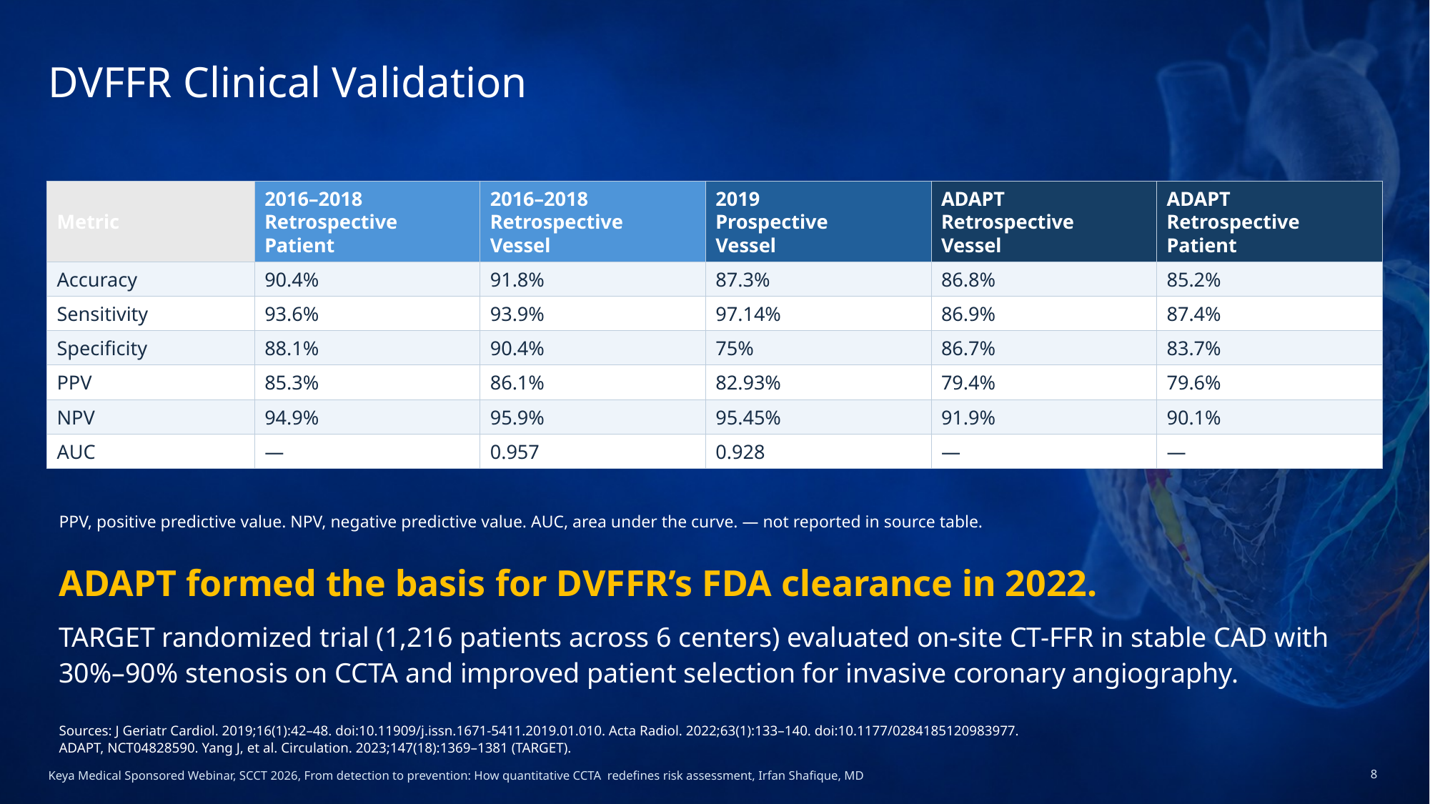
Clinical validation results presented for DEEPVESSEL FFR. Source: presentation by Irfan Shafique, MD.
Quantitative CT shows promise but standardization remains important
Dr. Zadeh examined the evidence from a deliberately critical perspective. Quantitative CT may improve the prediction of adverse cardiovascular outcomes, particularly through more precise measurement of total plaque burden. However, the evidence that quantitative analysis consistently outperforms expert interpretation or directly improves patient management remains incomplete.
He highlighted variability among commercial plaque-analysis platforms. The same lesion can produce substantially different measurements depending on the software, reconstruction approach, scanner, and analysis method. This variability is especially relevant when quantitative results are used for longitudinal follow-up. Consistent acquisition methods and, when possible, use of the same analysis platform are important for comparison over time.
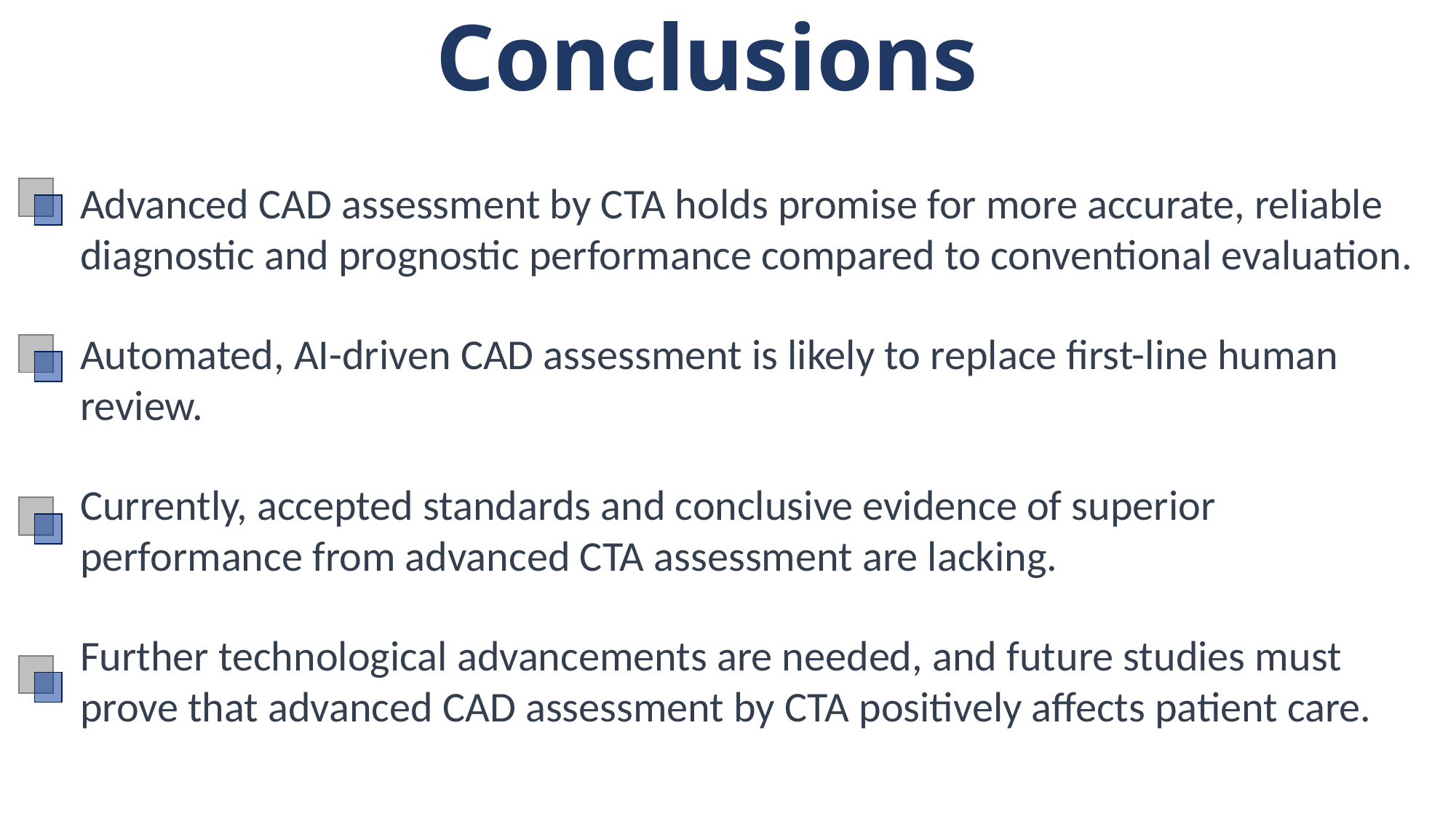
His central message was practical: AI-driven quantitative assessment is likely to play an increasingly important role, but clinicians still need accepted standards, stronger prospective evidence, and careful integration of quantitative outputs with the original images and the patient’s clinical context.
COMPOSE-CT validation of DEEPVESSEL Plaque
Dr. Budoff focused on the role of plaque imaging in prevention and presented the COMPOSE-CT clinical validation study for DEEPVESSEL Plaque. The multicenter study evaluated 147 analyzable CCTA examinations and 441 coronary vessels, comparing automated measurements with consensus assessments from three SCCT Level III readers.
At the patient level, the study reported strong correlations for total plaque volume (r=0.976), calcified plaque volume (r=0.994), and noncalcified plaque volume (r=0.945). CAD-RADS agreement was 75.5% for the exact category and 95.9% within one category, with a weighted kappa of 0.810. These results supported the FDA clearance of DEEPVESSEL Plaque in 2026.
Plaque analysis provides information that complements stenosis and CT-FFR. Total plaque burden describes the overall extent of atherosclerosis, while plaque composition can help distinguish calcified, noncalcified, and low-attenuation plaque. These findings may support a more individualized discussion of cardiovascular risk and preventive therapy.
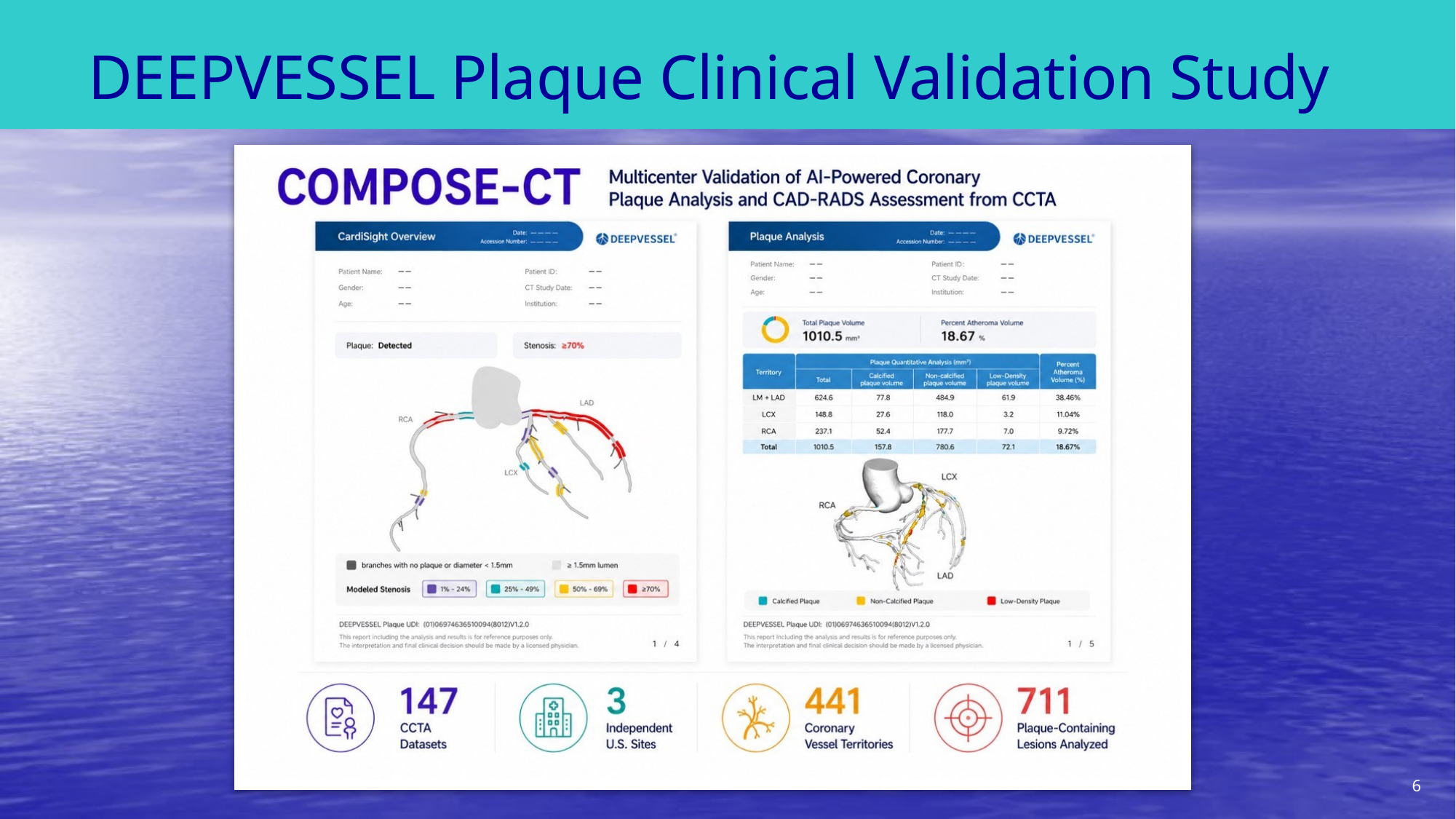
COMPOSE-CT multicenter clinical validation study overview. Source: presentation by Matthew J. Budoff, MD.
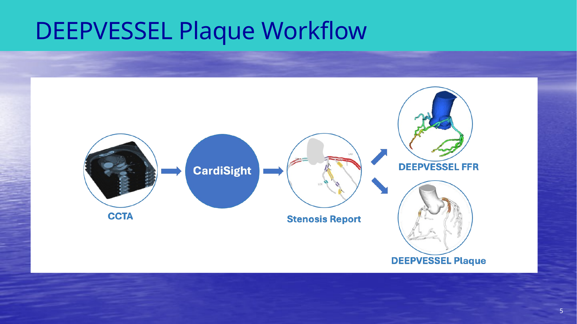
Comprehensive CCTA workflow with separate stenosis, plaque, and CT-FFR analyses. Source: presentation by Matthew J. Budoff, MD.
DVCloud: all analysis in one interactive viewer
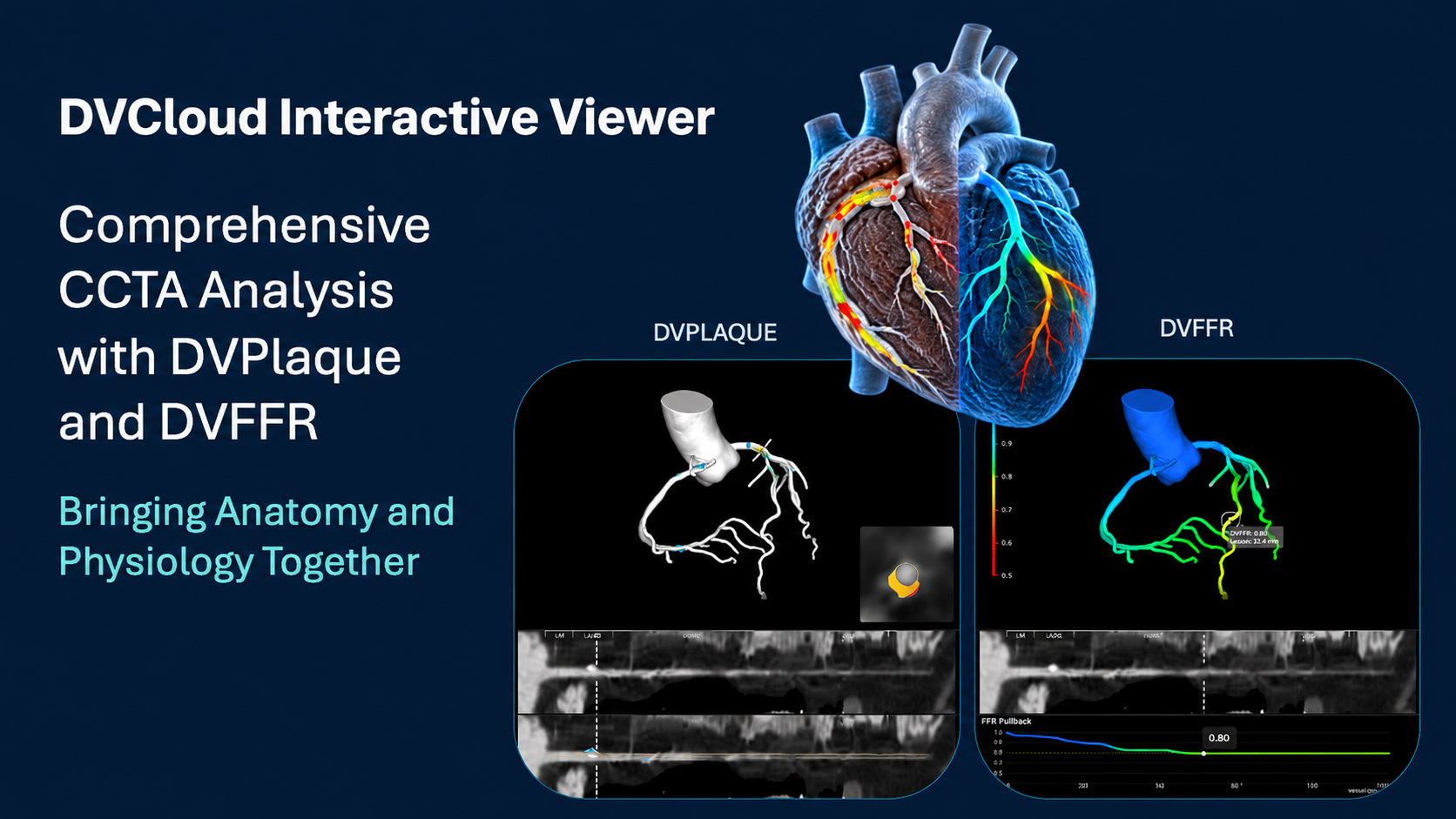
DVCloud brings stenosis, CT-FFR, and plaque analysis into one interactive environment.
Panel discussion
The faculty addressed several practical questions:
- Stents remain challenging because of metal artifacts and the difficulty of evaluating subtle in-stent restenosis, particularly in smaller stents.
- Perivascular fat attenuation and coronary inflammation are promising biomarkers, but the panel agreed that additional evidence is needed to establish how these measures should influence routine management.
- Serial plaque assessment should use consistent acquisition and analysis methods. Measurements from different vendors should not be assumed to be interchangeable.
- Quantitative plaque results are most useful as continuous measurements interpreted within the broader clinical picture, rather than reduced to a simple mild, moderate, or severe label.
- Education and communication with referring clinicians are essential if advanced imaging is to support earlier prevention rather than focus only on whether severe stenosis is present.